Anti-hnRNP-U / SAF-A antibody, rabbit serum (HUT)
$ 91.51
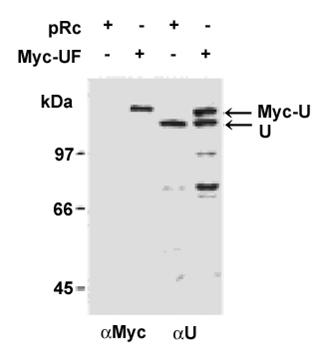
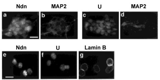
Description Heterogeneous nuclear ribonucleoprotein U (hnRNP-U, also known as scaffold attachment factor A, SAF-A) is a nuclear matrix-associated protein that interacts with chromosomal DNA. hnRNP-U specifically binds to scaffold/matrix attachment region of DNA and could thus be involved in higher order chromatin structure. hnRNP-U is also a RNA binding protein and forms complexes with heterogeneous nuclear RNA (hnRNA) and plays an important role in pre-mRNA processing and transport. HnRNP-U is reported to interact with necdin, a growth suppressor that is expressed in terminally differentiated neurons and skeletal muscle cells. It has been shown that hnRNP-U recruits necdin to the nuclear matrix where they form a stable complex. It is suggested that necdin suppresses cell proliferation through its interaction with hnRNP-U in the specific subnuclear structure (ref. 2). An antibody (named HUT) against mouse hnRNP-U was raised in rabbit (ref. 2). Applications Western blot (dilution: 1/3,000-1/1,000) Immunocytochemistry (dilution: 1/1,000-1/500) Immunoprecipitation Specification Immunogen: Recombinant MBT-fused mouse hnRNP-U (aa 614-800). Specificity: React with mouse and rat, and is estimated to react also with human from the amino acid sequence homology. Form: Antiserum added with 0.05% sodium azide Storage: Shipped at 4°C and stores at -20°C Data Link: Swiss-Prot Q8VEK3 (mouse), Q00839 (human) References: This antibody was described and used in ref. 2. Kiledjian M and Dreyfuss G (1992) “Primary structure and binding activity of the hnRNP U protein: binding RNA through RGG box.” EMBO J 11: 2655-2664 PMID: 1628625 Taniura H and Yoshikawa K (2002) “Necdin interacts with the ribonucleoprotein hnRNP U in the nuclear matrix.” J Cell Biochem 84:545-555 PMID: 11813259 Fig.1 Immunoblotting of hnRNP-U with this antibody (ref. 2). Specificity of anti-hnRNP-U antibody, HUT. Cell lysates were prepared from SAOS-2 cells transfected with pRc/CMV vectors (pRc) or pRc/CMV vectors expressing Myc-tagged hnRNP-U (Myc-UF). Exogenous Myc-tagged hnRNP-U (Myc-U) and endogenous hnRNP-U (U) proteins were detected by immunoblotting with anti-Myc antibody (αMyc) or HUT (αU). This antibody recognized exogenous Myc-tagged hnRNP-U and endogenous ~120 kDa hnRNP-U proteins in SAOS-2 cells. Fig.2 Immunocytochemistry using this antibody, HUT (ref. 2) Mouse P19 neurons were labeled with anti-necdin antibody (Ndn) (a) or with HUT for hnRNP-U (U) (c) in combination with anti-neuronal marker, MAP2, antibody for MAP2 (b, d). The nuclear matrix was prepared in situ and labeled for necdin (Ndn) (e), hnRNP-U (U) (f), and a nuclear matrix marker, lamin B (g). Both necdin and hnRNP-U were localized to the nuclei of differentiated neurons, which express the neuronal marker MAP2 (a-d). Necdin was also distributed in the neuronal cytoplasm (a). The immunocytochemical analysis of in situ extracted nuclear matrix revealed that both necdin and hnRNP-U were concentrated in intranuclear speckles throughout the nucleoplasm (e, f). Lamin B, a nuclear matrix marker, was localized to the nuclear lamina (g). These results suggest that both necdin and hnRNP-U are associated with the nuclear matrix of neurons.